Thermography
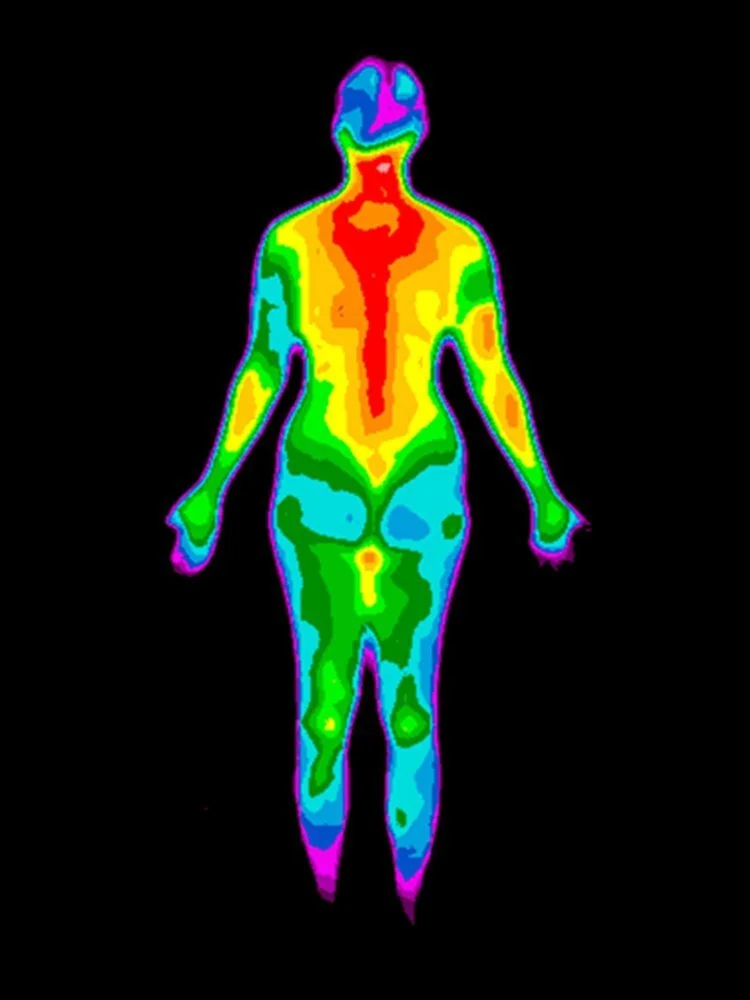
Thermography also known as thermal imaging is a non-contact and non-invasive procedure. The value of thermography as a screening tool is its ability to measure skin temperature changes. It offers individuals information that no other imaging technology can provide.
What is it?
Thermography captures and records temperature variations on the skin, which provides vital information directly influenced by complex metabolic and vascular activity. This information does not in any way suggest diagnosis and/or treatment. Studies show that the patient benefits when multiple tests are used together. This multimodal approach includes breast self-examinations, physical breast exams by a doctor, mammography, ultrasound, MRI, thermography, and other tests that may be ordered by your doctor. However, any suspicious finding will be accompanied with a strong and intentional recommendation for further clinical evaluation. If a lump or any other change in your breast is noticed before your next screening thermogram, consult your doctor immediately.
Thermal imaging is completely non-invasive and sought after for its ability to detect early changes where the body is prioritising inflammation. There is now a consensus that chronic inflammation is a clinical marker for a compromised immune system and the onset of chronic disease.
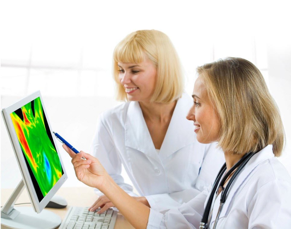
The very latest state-of-the-art medical grade Flir camera with total vision software, can detect the amount of thermal energy emitted by any area of your body. All images are interpreted by experts in clinical medical thermography, from which a full colour take-home report will enable you to visually see areas of inflammation present. Healthy lifestyle choices will be identified and applied during an in- depth follow-up naturopathic consultation, where you will be presented with your report.
What can thermography detect:
-
Description text goes here
-
Description text goes here
-
Description text goes here
-
Item description
Notice to patients presenting with previously diagnosed cancer:
Thermography interpretation in your report does not include information or recommendations related to the measured changes of disease beyond skin temperature changes and patterns. As there is no single known test capable of monitoring all biological influences of the complex disease generally diagnosed as cancer, continued monitoring with available additional testing as recommended by your personal physician is strongly advised. This thermal imaging health study will alerts you to the regions and conditions which may need further investigation including fibrocystic breasts, breast pain, blood perfusion, lymphatic congestion, dental issues, circulation vascular/heart disease, musculoskeletal problems, metabolic changes, digestive imbalance, diabetes detection.

Meet your Thermographer
Loulla Antoniou,
ARH, CMA, IAMT, NAANT
London’s most experienced Certified Clinical Thermographer, Loulla Antoniou, has joined our team at Hope Spring Clinic to carry out thermal imaging.
Loulla’s love and passion for holistic wellbeing is infectious. She is a Certified Clinical Medical Thermographer, with experience using the medical grade Flir thermal camera, and Total Vision™ software for our non- invasive thermal imaging.
Loulla’s natural ability to integrate the physical, mental, emotional, and spiritual is second nature, due to also being a registered licentiate homeopathic practitioner in homeopathic biopuncture, with over 20 years’ experience and post-graduate training. With background certification ranges from the USA, The Netherlands and United Kingdom, Loulla’s many years of training, experience and knowledge extends to bringing awareness to a healthy approach to beauty and aesthetics. Her preferred natural minimal treatment application as a certified aesthetic practitioner along with mesotherapy and sound medicine brings a fresh new perspective and approach.
What we offer
-
Full Body Study
The Upper Body Study involves taking 37 images of the entire body using thermography to detect inflammation, neovascular and neurological phenomena. Inflammation is a necessary response of the immune system to damage and infection. It signals the immune system to heal and repair damaged tissue and defend against foreign invaders such as viruses and bacteria. The imaging process takes approximately one hour.
Treatment time(s): 120 mins
Donations: £499
-

Breast Study
The Breast Study involves taking nine images of the breast, chest, and upper back, which takes about 30 minutes. Thermography can identify inflammatory, neovascular, and neurological issues based on temperature levels and patterns. A thermography study can also detect abnormal vascular patterns associated with chronic breast disease and monitor implant structural integrity.
Treatment time(s): 120 mins
Donations: £370
-

Womens Health Study
The Womens Health Study (Upper body) comprises 37 images of the entire body, including the head and neck, upper back, breast, abdomen and lower back, and trunk views. Imaging usually takes 1 hour. Thermography assists in identifying inflammatory, neovascular, and neurological phenomena based on the levels of temperature, differences in temperature, and appearance and location of thermal patterns.
Treatment time(s): 90 mins
Donations: £455
-

Mens Health Study
The Mens Health Study (Upper body) comprises 37 images of the entire body, including the head and neck, chest and upper back, abdomen and lower back, and trunk views. The imaging process typically lasts an hour. Thermography is used to detect inflammatory, neovascular, and neurological phenomena based on temperature levels, differences, and thermal patterns.
Treatment time(s): 90 mins
Donations: £455